Imaging and clinical correlation
A radiology report is a description of a picture, written by someone who never met the person in it.
Imaging is indispensable and routinely misused. It shows structure and reserve space. It does not show pain, it rarely dates an injury, and its findings appear at high rates in people with no symptoms at all — which is why a finding only becomes evidence once it is correlated to the examination.
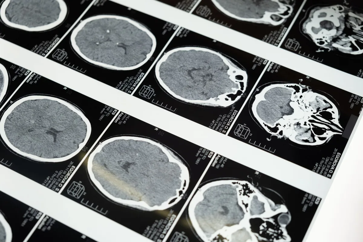
The base-rate problem
Brinjikji and colleagues pooled 33 studies of people without back pain: 37% of asymptomatic 20-year-olds show disc degeneration, rising to 96% by age 80; disc bulges rise from 30% to 84% across the same span. Boden reported the same phenomenon in 1990.
A finding that common cannot, by itself, distinguish a painful spine from a painless one. It is context, not conclusion.
What correlation actually means
- Does the level of the finding match the distribution of the symptoms?
- Does the side match?
- Do the examination findings — reflex, strength, sensation — map to the root the imaging implicates?
- If they do not, which is more likely wrong: the picture, or the person?
A reviewing report that recites findings without performing this correlation has skipped the step that converts an image into evidence, and that omission is visible on the face of the report.
Dating, honestly
Certain features suggest acuity, and a comparison with any prior study is worth more than all of them. A confident date derived from a single post-injury scan usually overstates what imaging supports, and we will not provide one.
Where no prior imaging exists, the honest statement is that the structural change is chronic and what changed is the symptom — which is the eggshell position, and a stronger one than a date nobody can defend.
This practice is not aligned to a side. Its obligation runs to the patient’s health and safety, which is the same reason it is not beholden to any position that would keep it from seeing what is actually there. We do not offer opinions on liability, we do not accept a referral or a review conditioned on reaching a particular conclusion, and we will say plainly when the evidence does not support the claim — whichever party was hoping otherwise.
Common questions
Should the films be sent, or just the report?
The films. The report is a summary written without the patient, and correlation cannot be done from it.
Can an MRI show pain?
No. It shows anatomy. Functional imaging research does not change this for individual clinical or forensic use.
What if imaging is normal but the patient is clearly injured?
Common. Facet-mediated pain, sacroiliac pain and many nerve injuries are invisible on MRI — see diagnostic nerve blocks.
Does a bigger herniation mean more pain?
No. Size correlates poorly with symptoms; position relative to the nerve root and the available reserve space matter more.
Related reading
- “Pre-existing degeneration”
- Objective corroboration
- Diagnostic nerve blocks
- Lumbar disc injury
- Causation and apportionment
Send the study, not the summary
We read the imaging against the examination and tell you whether the level of the finding matches the level of the symptom.
12166 Natural Bridge Rd
St. Louis, MO 63044
Monday to Friday, 8:00 a.m. to 5:00 p.m. Nothing on this page is legal advice, and nothing on it is medical advice.
Sources
- Brinjikji W et al. Systematic literature review of imaging features of spinal degeneration in asymptomatic populations. American Journal of Neuroradiology, 2015. PubMed 25430861
- Boden SD et al. Abnormal magnetic-resonance scans of the lumbar spine in asymptomatic subjects. Journal of Bone and Joint Surgery, 1990. PubMed 2312537
- Cohen SP et al. Consensus practice guidelines on interventions for lumbar facet joint pain from a multispecialty, international working group. Regional Anesthesia and Pain Medicine, 2020. PubMed 32245841
- Bono CM et al. An evidence-based clinical guideline for the diagnosis and treatment of cervical radiculopathy from degenerative disorders. The Spine Journal, 2011. PubMed 21168100