Sacroiliac joint pain · Diagnosis
A joint that cannot be diagnosed by examination or imaging, in a region every MRI is pointed at for other reasons.
Sacroiliac joint pain is a documented cause of low back and buttock pain that a lumbar MRI is not built to evaluate. It is diagnosed by image-guided block, and a 2025 multispecialty consensus guideline sets out how that should be done.
Why it is missed
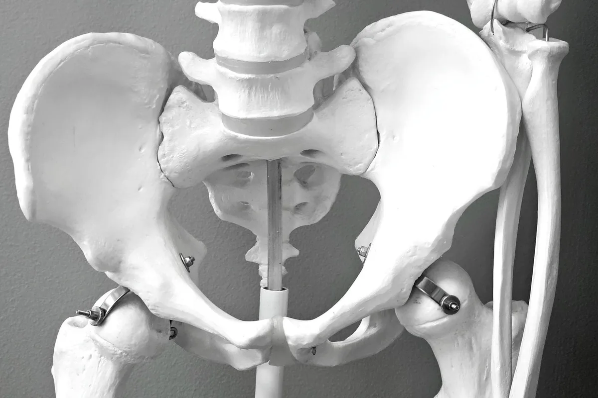
The joint sits deep behind muscle and bone. Physical examination provocation tests have limited individual accuracy, and lumbar imaging is directed at discs and canal rather than at the joint. So a client with genuine sacroiliac pain accumulates normal studies, which is then read as absence of injury.
McCormick and colleagues published consensus practice guidelines on sacroiliac joint complex pain from a multispecialty international working group in 2025; Szadek and colleagues provide a practice review. Both address the central point: the diagnosis rests on image-guided diagnostic blocks rather than on imaging or examination alone.
What corroborates it
An image-guided block of the joint or its lateral branch supply, performed with fluoroscopic confirmation and recorded against timed measurements — the same methodology as any other diagnostic block, and repeated before anything irreversible.
The evidence tier for sacroiliac orthobiologic injection specifically is lower than for the knee, and ASIPP grades sacroiliac PRP at Level IV with a low consensus recommendation. That is stated at its level here — see the evidence by indication.
In an injury case
Common mechanisms include a fall onto one buttock, a lateral impact, and the pelvic loading of a braced leg on a brake pedal at the moment of collision. Postpartum and post-fusion presentations exist and predate many claims, which makes the pre-injury record important rather than fatal — see causation and apportionment.
This practice is not aligned to a side. Its obligation runs to the patient’s health and safety, which is the same reason it is not beholden to any position that would keep it from seeing what is actually there. We do not offer opinions on liability, we do not accept a referral or a review conditioned on reaching a particular conclusion, and we will say plainly when the evidence does not support the claim — whichever party was hoping otherwise.
Common questions
Why does the MRI not show it?
A lumbar MRI is aimed at the discs and canal, and degenerative change at the joint is common and poorly correlated with pain. The diagnosis is made by block.
Are examination provocation tests enough?
Individually they are limited. Clusters perform better and still do not replace a diagnostic block.
Can a fall cause it?
A fall onto one buttock is a classic mechanism, as is asymmetric loading in a collision.
What treatment follows a positive block?
It depends on the response and the rest of the picture. A positive block establishes the target; it does not dictate one treatment.
Related reading
- Diagnostic nerve blocks
- Lumbar disc injury
- Imaging and correlation
- What the evidence says
- Causation and apportionment
Ask whether the joint was ever actually tested
In most files with buttock pain and normal imaging, it was not.
12166 Natural Bridge Rd
St. Louis, MO 63044
Monday to Friday, 8:00 a.m. to 5:00 p.m. Nothing on this page is legal advice, and nothing on it is medical advice.
Sources
- McCormick ZL et al. Consensus practice guidelines on sacroiliac joint complex pain from a multispecialty international working group. Pain Medicine, 2025. PubMed 41318933
- Szadek K et al. Sacroiliac joint pain. Pain Practice, 2024. PubMed 38155419
- Cohen SP et al. Multicenter, randomized, comparative cost-effectiveness study comparing 0, 1, and 2 diagnostic medial branch block treatment paradigms before lumbar facet radiofrequency denervation. Anesthesiology, 2010. PubMed 20613471
- Brinjikji W et al. Systematic literature review of imaging features of spinal degeneration in asymptomatic populations. American Journal of Neuroradiology, 2015. PubMed 25430861