Pre-existing degeneration on MRI
Degenerative changes are so common in people with no pain at all that finding them tells you almost nothing about why the patient hurts.
Pre-existing degeneration on an MRI rarely explains why a patient hurts. Pooled data from 33 studies found disc degeneration in 37% of pain-free 20-year-olds and 96% by age 80, so a degenerative finding is a base rate, not a diagnosis.
The most reliable defense argument in spine litigation is a radiology report. It is also the weakest, because the same findings appear at high rates in volunteers who have never had back pain in their lives — and that literature is thirty-five years old, uncontroversial, and rarely put in front of a jury.
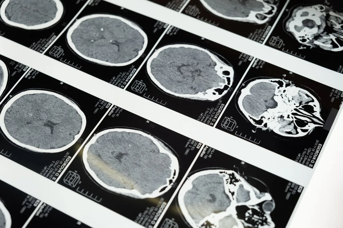
How common is disc degeneration in people with no back pain?
In 2015 Brinjikji and colleagues pooled 33 studies of people with no back pain and reported the age-specific prevalence of exactly the findings that populate a reviewing expert’s report.
37%
of asymptomatic 20-year-olds already show disc degeneration. By age 80 it is 96%. Disc bulges rise from 30% to 84% across the same span — all in people with no pain.
Boden had shown the same thing in 1990 with a smaller sample and a blunter conclusion: roughly a third of asymptomatic subjects had a substantial abnormality on lumbar MRI, and the rate climbed steeply with age.
The implication is not that MRI is useless. It is that a degenerative finding is a base rate, not a diagnosis. Reporting that a 52-year-old has multilevel disc desiccation is close to reporting that they have gray hair.
Does pre-existing degeneration explain the pain after an accident?
The argument is built in two moves. First, the report establishes that degeneration exists. Second, the expert asserts that the degeneration, rather than the collision, explains the pain. The first move is unobjectionable. The second one is where it fails.
BREAK ONE — THE BASE RATE
If the finding is present in most pain-free people of the same age, its presence cannot distinguish a painful spine from a painless one. An expert who treats it as causal has to explain why the finding was silent for fifty-two years and became the whole explanation in the month after a collision.
BREAK TWO — NO CORRELATION TO THE EXAM
Degeneration at L2-3 does not explain pain in an S1 distribution. A reviewing expert relying on the report often has not correlated the level of the finding with the level of the symptoms, because doing so frequently destroys the argument. That correlation is ordinary clinical work — see imaging and correlation.
Can an accident aggravate pre-existing degeneration?
There is a version of the degeneration finding that helps the patient, and it is not a spin. A degenerated segment has less reserve: a narrowed foramen, a stiffened motion segment, a facet joint already carrying load it was not designed for. Add a traumatic force and the margin that a healthy spine would have absorbed is not there.
That is the eggshell argument made in anatomical terms, and it is the honest reading of the same film both sides are holding. The argument runs: the degeneration explains the pain. The correct answer is that the degeneration explains why this force produced this much pain in this person.
What converts that from argument to evidence is a test that names the structure. A degenerated facet joint that is generating pain can be identified with medial branch blocks; multi-society guidelines treat a concordant response on blocks as the reference standard for facet-mediated pain precisely because imaging cannot make that call.
What we will not say
We will not testify that a degenerative finding was caused by a collision when the imaging shows a chronic process, and we will not date degeneration to an accident from a single post-injury study. Where a comparison film exists, it is read. Where it does not, the honest statement is that the structural change is chronic and the symptom is what changed.
This practice is not aligned to a side. Its obligation runs to the patient’s health and safety, which is the same reason it is not beholden to any position that would keep it from seeing what is actually there. We do not offer opinions on liability, we do not accept a referral or a review conditioned on reaching a particular conclusion, and we will say plainly when the evidence does not support the claim — whichever party was hoping otherwise.
Common questions
Can an MRI date a disc herniation?
Rarely with precision. Certain features suggest acuity, but a confident date from one post-injury study usually overstates what imaging supports. Comparison with any prior study is worth far more — see imaging and correlation.
If degeneration is that common, is imaging worth ordering?
Yes, for what it rules out and for surgical planning. It is a poor tool for attributing a symptom to a structure, which is what diagnostic blocks exist to do.
The client had prior back complaints. Is the case gone?
No. Prior complaints change the question from causation to aggravation, which is a question a treating physician can address with a documented change in pattern, intensity and function.
What if the reviewing expert cites the same asymptomatic literature?
They should. It cuts against confident causal claims in both directions, which is why the case should not rest on imaging at all but on objective corroboration of a named generator.
What is considered a pre-existing injury?
In a spine claim, the pre-existing finding is usually degeneration on an MRI: disc desiccation, disc bulges, a narrowed foramen, or a facet joint already carrying extra load. Prior back complaints count too. Neither one settles the case. Degeneration shows up in most pain-free people as they age, and prior complaints change the question from causation to aggravation.
Can a car accident cause degenerative disc disease?
Degeneration is a chronic process. We will not testify that a collision caused a degenerative finding when the imaging shows a chronic change, and we will not date degeneration to an accident from a single post-injury MRI. Where no earlier film exists, the honest statement is that the structural change is chronic and the symptom is what changed. A degenerated segment has less reserve, which is why the same force can produce more pain.
Related reading
- The eggshell plaintiff
- Imaging and correlation
- Diagnostic nerve blocks
- Causation and apportionment
- Objective corroboration
Send the films, not just the reports
A radiology report is a summary written without the patient in the room. We read the study against the exam and tell you whether the level of the finding matches the level of the symptom.
12166 Natural Bridge Rd
St. Louis, MO 63044
Monday to Friday, 8:00 a.m. to 5:00 p.m. Nothing on this page is legal advice, and nothing on it is medical advice.
Sources
- Brinjikji W et al. Systematic literature review of imaging features of spinal degeneration in asymptomatic populations. American Journal of Neuroradiology, 2015. PubMed 25430861
- Boden SD et al. Abnormal magnetic-resonance scans of the lumbar spine in asymptomatic subjects. Journal of Bone and Joint Surgery, 1990. PubMed 2312537
- Cohen SP et al. Consensus practice guidelines on interventions for lumbar facet joint pain from a multispecialty, international working group. Regional Anesthesia and Pain Medicine, 2020. PubMed 32245841
- Cohen SP et al. Multicenter, randomized, comparative cost-effectiveness study comparing 0, 1, and 2 diagnostic medial branch block treatment paradigms before lumbar facet radiofrequency denervation. Anesthesiology, 2010. PubMed 20613471